Contents
Idiopathic retinal vascular disorder with retinal telangiectasia with intraretinal and/or subretinal exudation without appreciable retinal or vitreal traction.
History:
George Coats first described this condition in 1908 as unilateral retinal vascular abnormality with exudation occurring in young males. Subsequently, Leber reported another entity characterized by multiple retinal aneurysms, associated with retinal degeneration. In 1955, Reese described the similarities between Coats disease and Leber miliary aneurysms and concluded that they represent a spectrum of the same disease with the underlying pathology being retinal telangiectasia leading to progressive exudation and retinal detachment.
Pathophysiology
Breakdown of blood retinal barrier (BRB):
Due to changes at the endothelial level of the retinal vasculature.
Plasma leaks into the vessel wall resulting in thickening and “sausage-like” shape of vessels.
Presence of abnormal pericytes
Abnormal pericytes, which along with the damaged endothelium causes bulging of the vessels and the characteristic telangiectasia
The damaged vasculature often rests in a bed of relative retinal ischemia. Exudation of lipids from these vessels is responsible for retinal detachment, retinal thickening, and retinal cyst formation.
Presentation
Unilateral involvement in young males is the typical presentation with most cases being diagnosed in the first and second decade of life. Younger the patient, more severe is the presentation and poorer the visual outcome.
Retinal vascular features:
- Vision loss
- Strabismus
- Xanthocoria
- Nystagmus
- Pain
Anterior segment findings:
- Corneal edema, megalocornea, shallow anterior chamber, anterior chamber cholesterolosis, neovascularization of iris and resultant iris heterochromia, and cataract.
Complications:
Most of the complications are secondary to chronic retinal detachment.
- Neovascular glaucoma with iris and angle neovascularization
- Anterior chamber cholesterolosis: Result of migration of cholesterol crystals from exudates in subretinal space into anterior chamber
- Retinal and vitreous hemorrhage (3% cases)
- Retinal macrocysts (degenerative changes)
- Vasoproliferative tumor (vascular mass in the fundus developing in response to chronic retinal detachment)
Diagnosis
Fundus examination:
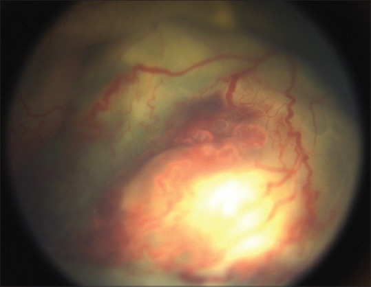
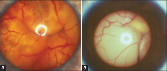
- “Light bulb telangiectasias” because of the bulbous terminal configuration and associated extensive yellow exudation
- Retinal detachment
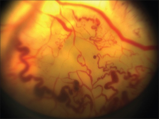
Fluorescein angiography (FFA):
- Idiopathic retinal telangiectasia as a ‘light bulb appearance‘
- Peripheral nonperfusion
- Other features: Irregular retinal vessel dilatations, retinal vessel tortuosity, areas of capillary non-perfusion, intra- and/or subretinal exudation and retinal detachment

Differential diagnosis:
- Retinoblastoma
- Toxocariasis
- Persistent fetal vasculature syndrome
- Retinitis pigmentosa with Coats-like reaction
- Diseases with vascular changes at the vitreoretinal interface (retinopathy of prematurity, familial exudative vitreoretinopathy, incontinentia pigmenti)
Management
Aim of management is to address and eradicate the telangiectatic vessels in order to control and cause resolution of intra- and subretinal exudates and ultimately salvage the globe with preservation of vision.
- Cryotherapy
- Laser photocoagulation
- Anti-VEGF therapy