
Contents
Acute highly contagious respiratory tract infection, caused by Bordetella pertussis.
- Aka, 100 Day Cough
History:
In the seventh century, during the Sui Dynasty, a pertussis-like illness was described by Chinese medical scholar Yuanfang Chao as “the cough of 100 days”. One thousand years later (1578) in France, Guillaume De Baillou provided the first description of whooping cough among children of Paris; he described the illness as “quinte” due to the observed 5-h periodicity of paroxysms seen in acute episodes of disease. Recent research suggests that the earliest recorded epidemics of pertussis were noted in Persia (present-day Iran). In Europe, pertussis outbreaks were first described in the 16th century, but recognition of the causative agent did not occur until 300 years later. In 1883, a German scientist, Carl Burger, working at the University of Bonn recognized rods of bacteria in a stained sputum specimen from a patient with clinical pertussis. Seventeen years later, Jules Bordet, a Belgian physician-scientist, visualized small Gram-negative bacilli in the sputum of his 5-month-old daughter suffering from whooping cough. However, at the time, Bordet was unable to grow these bacilli in available culture media. Six years later, Bordet’s son had also suffered from pertussis, and by that time, Bordet and Gengou were successful in isolating and growing B. pertussis for the first time in history. In 1920, Bordet was awarded the 1919 Nobel Prize in Physiology or Medicine for his work related to antimicrobial immunology, which included extensive study of B. pertussis and set the stage for identification of this organism as the cause of whooping cough.
Epidemiology
- Highly contagious (100% attack rate))
- Transmission: Aerosol droplets
- Reservoir & source: Coughing adolescents & adults
- Subclinical infection (80%)
- In fully immunised or naturally immunised patients
Etiology
Causative organisms:
- Bordetella pertussis
- B. parapertussis
- B. brochiseptica
- Adenovirus (1, 2, 3 & 5)
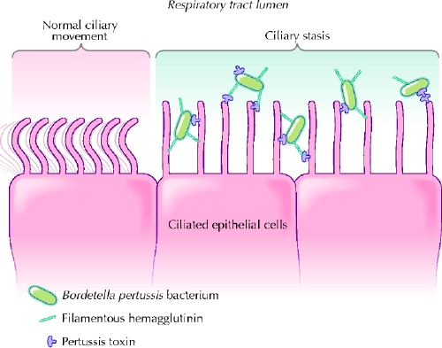
Pathophysiology
Ciliary stasis:

Presentation
After an incubation period of 1 to 3 weeks, pertussis infection typically progresses through three distinct stages: the catarrhal phase, the paroxysmal phase, and the convalescent phase.
I. Catarrhal stage: 1-2 weeks
Similarly to other upper respiratory tract infections
- M/infectious period
- Cough & coryza with little nasopharyngeal secretions
- Progressing severity
- Paroxysmal in early stages
- Frequent during night
II. Paroxysmal stage: 2-6 weeks
Characterized by paroxysms of a staccato cough and resolution of fever.
- Cough
- Episodic paroxysms (Precipitated by food, cold air & cold liquids)
- Increasing intensity
- Thick tenacious mucus
- Inspiratory whoop “whooping cough”
- High pitched
- Due to air rushing in during inspiration through half-open glottis
- Associated vomiting
- Ulceration (frenulum of the tongue)
- Repeated thrusting of tongue over the teeth
III. Convalescent stage: 1-4 weeks
Residual cough persists for weeks to months
- General improvement:
- ↓ Intensity & paroxysms of cough
- ↓ Vomiting
Complications
Respiratory complications:
- Otitis media
- Pneumonia
- Atelectasis
- Emphysema
- Bronchiectasis
- Pneumothorax
- Pneumomediastinum
Neurological complications:
- Seizures & Encephalopathy (2-7%)
Bleeding episodes:
- Epistaxis
- Retinal/subconjunctival bleeds
- Intracranial haemorrhage
Mechanical complications:
- Inguinal hernia, rectal prolapse
Malnutrition
Due to persistent vomiting
Flare-up of tuberculosis
Diagnosis
Lab investigations:
- Lymphocytic leucocytosis
- ↓ ESR
Chest x-ray:
Findings are nonspecific and may show peribronchial thickening, atelectasis, or infiltrate
- ‘Shaggy’ right heart border (CHARACTERISTIC)
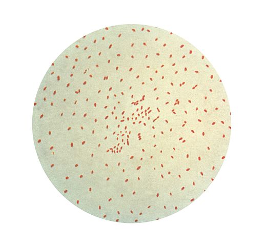
PCR or nasopharyngeal culture:
The fastidious and slow-growing Bordetella organisms require specialized media and cultures are typically not positive for 3 to 7 days. In adults, by the time the diagnosis is suspected, cultures are typically negative (96%), and overall culture sensitivity is only 20% to 40%.
Management
Supportive management:
- Oxygen
- Suctioning
- Hydration
- Avoidance of respiratory irritants
Hospitalization:
Indicated for patients with superimposed pneumonia, hypoxia, CNS complications, or who are unable to tolerate nutrition and hydration by mouth.
- Antibiotic therapy: Erythromycin (DOC, also used in chemoprophylaxis of contacts)
- Alternatives: Azithromycin, clarithromycin
Summary
One reply on “Pertussis”
[…] (M/C): Post-pneumonia, whooping cough, measles, mycobacterial […]