untitled
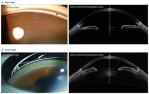
Gonioscopic Imaging and Optical Coherence Tomographic Imaging of Open-Angle and Closed-Angle: A lens with a prism is placed on the eye during gonioscopy, a process during which the examiner is able to examine the angle configuration and assess for the presence of angle closure. A, The arrowhead points to the lack of contact between the iris and angle. Image on the right shows the anterior segment captured by optical coherence tomography. The arrowheads point to visible trabecular meshwork. B, The angle is closed with the trabecular meshwork not visible due to apposition of the iris to the angle. In the right image, the arrowheads indicate apposition of the iris to the angle wall; the anterior chamber is shallow and the iris has a slightly convex configuration. This is more noticeable in the region of the iris on the right. | Weinreb, R. N., Aung, T., & Medeiros, F. A. (2014). The pathophysiology and treatment of glaucoma: a review. JAMA, 311(18), 1901–1911. https://doi.org/10.1001/jama.2014.3192
